The 'plica syndrome' is a group of symptoms that are commonly associated with the presence of an abnormally thickened plica.
A plica is a fold of the soft inner lining of the knee joint (this lining is called the 'synovium', so a plica is also often called a 'synovial plica'). Plicae (plural) are remnants of structures which existed in the foetus (in the womb) and which subsequently fell away to a lesser or greater extent as the knee developed. In some people they are still significant stuctures and in other people they may be completely absent.
Symptoms of the plica syndrome will depend upon which plica is causing the problem. A problematic medial plica may have the following associated with it:
- pain along the inner aspect of the patella
- a string-like cord under the skin in this area
- intermittent 'catching' of the joint at a particular point in extension
- occasional 'pseudo'-locking, ie the joint seems to lock but can be easily unlocked
- stiffness and pain after sitting still for a long time, eg at a movie
- occasional giving-way
- Typically plicae are found in three positions -
- suprapatellar (SPP) (above the patella or kneecap)
- infrapatellar (IPP) (below the patella), sometimes called the 'ligamentum mucosum' or 'filmyligament' as it mimics the cruciate ligament inside the knee, but is not a ligament at all.
- medial (MPP) (on the inner aspect of the knee). It may form a shelf-like band and is then referred to as a 'medial shelf'.
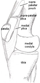
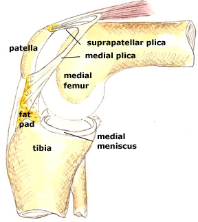
In the illustration on the left, looking at the knee bones of the right knee from its inner aspect, you can see the relationship of the media and suprapatellar plica. Here the leg is straight.
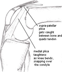
When the knee is bent (see image on the right), the plicae taughten, and a medial plica can snap abruptly over the medial femoral condyle, while the suprapatellar plica may be nipped between condyle and the quadriceps tendon.
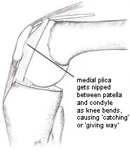
The medial plica can also become nipped (see left) between patella and condyle, causing momentary pain and 'giving way'.
A damaged plica may settle on its own, or respond to non-surgical measures such as physiotherapy or injections. However, sometimes the nipping continues and the joint cartilage becomes damaged and arthritis may set off. In this case surgery to remove the plica is the best option.
No comments:
Post a Comment